
Apoptosis: "In Situ" Identification by Nuclei DNA Fragments-Specific Tagging, in Endomyocardial Biopsyes During Episodes of Acute Rejection
pp 241-247
DOI:
https://doi.org/10.7775/rac.v63i3.3580Keywords:
Apoptosis, Acute rejection, Transplant , Endomyocardial biopsyAbstract
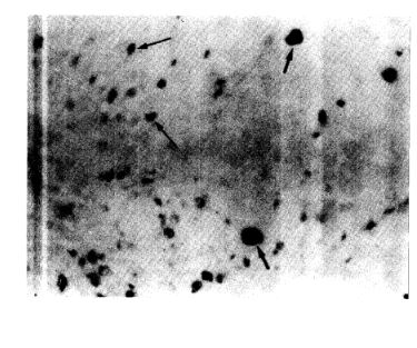
The presence of apoptosis in individual nuclei of endomyocardial biopsies from 6 patients with allogeneic heart transplant was demonstrated by means of the detection in situ of nuclear DNA breaks. Biotinylated dUTP incorporated with deoxynucleotidyl transferase and revealed with alkaline phosphatase-labeled streptavidin was employed. None of the samples at the moment of transplant presented apoptotic nuclei, which were observed in the myocytes, endothelial and connective tissue interstitial cells of the biopsies, obtained between 7 and 146 days after transplant. The kind and number of cells with apoptotic nuclei was different according to the rejection grade (Grading of the ISHLT). All the rejection grade 3A (n = 8),half of the rejection grade 2 (n = 8) and some of the rejection grade 1B (3/8) biopsies showed myocytes with apoptotic nuclei within or in the neighborhood of the inflammatory infiltrates zones. In therejection grades 0 and 1A, and in "Quilty" effect areas, no myocytes with apoptotic nuclei were observed. On the contrary, in the endothelial and connective tissue cells, apoptotic nuclei were observed in all the rejection grades. These results indicate that during the acute rejection episodes myocyte apoptosis is one of the mechanisms of immunological damage and that its recognition may represent a useful tool for the demonstration of myocyte damage in endomyocardial biopsies.
Downloads
Published
Issue
Section
License
Copyright (c) 2026 Argentine Journal of Cardiology

This work is licensed under a Creative Commons Attribution-NonCommercial-ShareAlike 4.0 International License.


