
Efficacy and safety of dual-axis rotational coronary angiography versus conventional angiography
pp. 280-285
DOI:
https://doi.org/10.7775/rac.es.v80.i4.1489Keywords:
Rotational Angiography, Coronary Artery Disease, RadiationAbstract
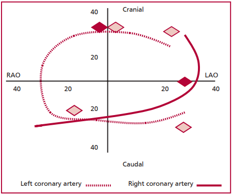
Background: Conventional coronary angiography (CA) is the gold standard for the diagnosis of coronary artery disease. However, this technique requires several orthogonal projections to determine the severity of the disease. Dual-axis rotational coronary angiography (DARCA) is a new technique which acquires the image of each coronary artery using a single contrast injection, potentially reducing both radiation and contrast exposure.
Objective: The aim of this study was to determine the amount of contrast used, radiation exposure and diagnostic accuracy of DARCA compared to conventional CA.
Methods: We conducted a prospective, self-controlled study of consecutive patients undergoing elective coronary angiography to compare DARCA versus the conventional technique. All the angiographies were reviewed by two independent interventional cardiologists (observer 1 and observer 2) who evaluated agreement between both types of images. The observers evaluated firstly DARCA and three weeks later conventional CA images to prevent bias in their interpretation.
Results: The contrast volume used in the diagnostic procedure was significantly lower with DARCA (33.29 ± 11.2 ml vs. 17±5.4 ml; p < 0.01). Radiation exposure in the patient (235.6 ± 76.8 mGy vs. 82.7 ± 46.6 mGy; p <0.01) and operator was also significantly lower (5.7 ± 3.26 µSv vs. 2.48 ± 1.47 µSv; p <0.04). A slight difference and a strong correlation were seen in the number of lesions, vessel diameter and percentage of stenosis between both observers and both methods. Categorical assessment of percentage of stenosis also showed adequate agreement.
Conclusion: DARCA reduces the use of contrast agents and radiation exposure compared to the conventional technique without modifying the diagnostic accuracy of the method.
Downloads
Published
Issue
Section
License
Copyright (c) 2025 Argentine Journal of Cardiology

This work is licensed under a Creative Commons Attribution-NonCommercial-ShareAlike 4.0 International License.


