
Electrocardiography During the Third Trimester of Pregnancy: Description of its Characteristics
pp 131-135
DOI:
https://doi.org/10.7775/rac.es.v89.i2.20011Keywords:
Pregnancy Complications, Cardiovascular, Pregnancy Trimester, Third, Electrocardiography, Cardiovascular diseaseAbstract
Objective: The aim of this study was to analyze the electrocardiographic characteristics in pregnant women without cardiovascular
disease.
Methods: This was a descriptive, cross-sectional, multicenter study, including patients without cardiovascular disease in their third
trimester of pregnancy, who underwent cardiac evaluation before delivery between April and July 2020. All patients signed the corresponding informed consent.
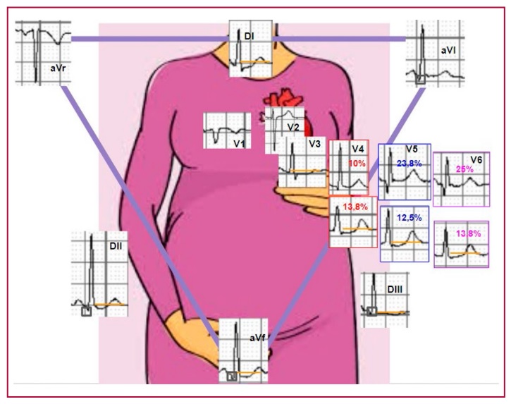
Results: A total of 80 tracings were analyzed. Median heart rate was 82 bpm (IQR 70-93 bpm) and median QRS axis was 54° (IQR
39°-71°). Q waves and ST-segment depression were observed in the inferior wall and from V4 to V6. Median QTc was 422 msec (IQR
404-445 msec) and median time from T-peak to T-end was 86 msec (IQR 74-95 msec).
Conclusion: The most common changes occurred in leads III and II, aVF and from V4 to V6. Main changes included Q waves and
ST-segment depression. Axis deviations, sinus tachycardia or prolonged QTc were rare.
Downloads
Published
Issue
Section
License
Copyright (c) 2025 Argentine Journal of Cardiology

This work is licensed under a Creative Commons Attribution-NonCommercial-ShareAlike 4.0 International License.


