
Evaluation of the Diagnostic Capacity of the Electrocardiagram in Ventricular Hypertrophy
pp 357-366
DOI:
https://doi.org/10.7775/rac.v62i4.3533Keywords:
Ventricular hypertrophy, Electrocardiogram, Pathologic findingsAbstract
Background
Ventricular hypertrophy is a frequent heart condition and numerous electrocardiographic criteria have been described for its diagnosis without a completely satisfactory correlation.The objectives of this study were: 1) To establish more accurate electrocardiographic criteria for the diagnosis of ventricular hypertrophy. 2) To correlate the degree of ventricular hypertrophy according to the pathologic findings with the number of criteria met.
Methods
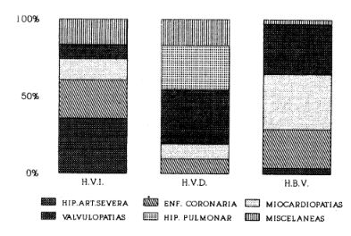
One hundred and two patients who died in our institution between 1981 and 1991 were studied. Ventricular hypertrophy was defined as heart weight >450 grams for male subjects and > 400 grams for female subjects. Sixty-eight patients presented left ventricular hypertrophy (left ventricle free wall and/or septal thick-ness > 1.9 cm and hypertrophy of the myocardial fibers with "bizarre" nuclei); 21 patients presented right ventricular hypertrophy (right ventricular free wall thickness > 0.7 cm) and 28 presented biventricular hypertrophy.The remaining 25 subjects constituted the control group(absence of ventricular hypertrophy or heart disease in the pathologic findings). The sensitivity and specificity of the electrocardiogram was assessed for the diagnosis of left ventricular hypertrophy by means of using 23 criteria and 4 methods; 21 criteria and 2 methods for right ventricular hypertrophy and additional 44 electrocardiogaphic criteria for biventricular hypertrophy.
Results
When the free wall thickness of each ventricle was correlated with the number of electrocardiographic criteria met, asymmetric septal hypertrophy with a septa)thickness < 2.4 cm presented few electrocardiographic signs of left ventricular hypertrophy (0 to5) and left ventricular hypertrophy with a free wall thickness >2.4 cm as well as right ventricular hypertrophy with a free wall thickness < 0.9cm did not present a direct relationship with free wall thickness and the number of electrocardiographic criteria met for hypertrophy. The more sensitive criteria for the diagnosis of left ventricular hypertrophy were:ST-segment depression of DI, aVL,V5 and V6 (64%); incomplete left bundle branch block(57%) and left atria] hypertrophy (53%). Pathologic Q waves were present only in 21.7% of the subjects with asymmetric septal hypertrophy.The most sensitive criteria for the diagnosis of right ventricular hypertrophy during electrocardiogram were: R/S in V 1 > 1 with R waves > 5mm (60%) and incomplete right bundle branch block with high sensitivity (81%) but with(70%) specificity. In the case of biventricular hypertrophy we observed high voltage and widened complexes in the precordial.~ lead which met the criteria of right ventricular and/or left ventricular hypertrophy. In the case of biventricular hypertrophy, the best diagnostic criteria for left ventricular hypertrophy was S wave in V2 + R wave inV5 > 35 tam (60%) and the most sensitive for right ventricular hypertrophy was incomplete right bundle branch block (42%).
Conclusions
Electrocardiogram presented a low sensitivity and a high specificity in the diagnosis of ventricular hypertrophy. Only in the case of severe hypertrophies, there was a direct relation between the degree of hypertrophy and the number of electrocardiographic criteria met.
Downloads
Published
Issue
Section
License
Copyright (c) 2026 Argentine Journal of Cardiology

This work is licensed under a Creative Commons Attribution-NonCommercial-ShareAlike 4.0 International License.


