Dilated cardiomyopathy induces electrical remodelingin the human ventricle
pp 109-115
DOI:
https://doi.org/10.7775/rac.v72i2.2856Keywords:
Electrophysiology, Cardiomyopathy, RemodelingAbstract
Background: Previous studies have shown marked heterogeneity acrossthe ventricular wall. Epicardial (EPI), endocardial (ENDO) and M cells differ in their repolarization characteristics: EPI nd M cells display a prominent “spike and dome” morphology, whereas M cells display a more prolonged action potential duration, especially at slow rates. Prolonged action potentials have been observed in the hearts of patients with terminal heart failure.
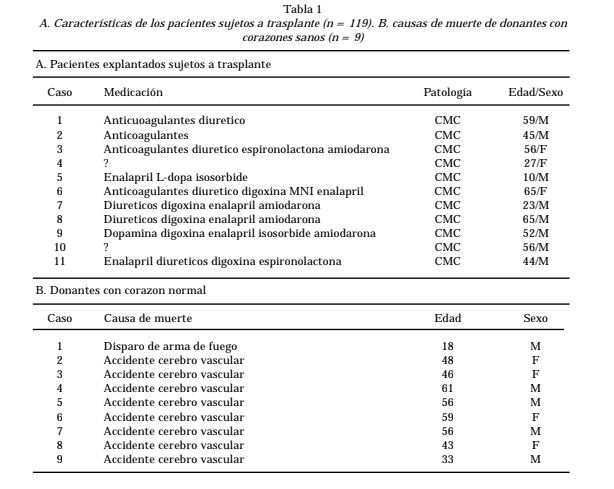
Research design and methods: This study used microelectrode techniques to record action potentials from EPI, M region, and ENDO tissues isolated from the apex of 11 explanted hearts with dilated cardiomyopathy (DCM) as well as from 9 normal hearts.
Results: In normal hearts, M cell action potential durations (APD)were significantly longer than those of EPI or ENDO, especially at slow rates (basic cycle length, BCL= 5000 msec; APD90 = 271+10 , 346+12 and 238+9 msec in EPI, M andENDO, respectively). In DCM hearts, ENDO and EPI APD were significantly increased but remained relatively un-changed in M cells (APD90 = 336+17, 351+17 and 339+16 msec in EPI, M and ENDO, respectively). Spike and dome morphology of EPI and M cells was attenuated in DCM hearts. The class III agent d-sotalol induced a preferential prolongation of M cell APD in both normal and DCM hearts(n = 3).
Conclusion: DCM induces electrical remodeling by altering the morphology of action potentials of epicardial, endocardial and M cells.
Downloads
Published
Issue
Section
License
Copyright (c) 2026 Argentine Journal of Cardiology

This work is licensed under a Creative Commons Attribution-NonCommercial-ShareAlike 4.0 International License.