Two-dimensional Strain Echocardiography in the Evaluation of Ventricular Function in Patients with Newly Diagnosed Hereditary Hemochromatosis
pp. 429-435
DOI:
https://doi.org/10.7775/rac.v87i6.801Keywords:
Echocardiography - Hemochromatosis - Heart Diseases - IronAbstract
Background: Hereditary hemochromatosis is a genetic disorder characterized by systemic iron overload which can involve the heart in advanced stages. Early myocardial involvement has not been thoroughly investigated.
Objective: The aim of our study was to evaluate the performance of conventional Doppler-echocardiography and 2D strain echocar diography in a group of patients with newly diagnosed hereditary hemochromatosis vs. a control group.
Methods: Patients with newly diagnosed hereditary hemochromatosis without treatment were included. All the patients underwent conventional Doppler echocardiography and 2D strain echocardiography with evaluation of left ventricular longitudinal, radial and circumferential strain, twist and torsion.
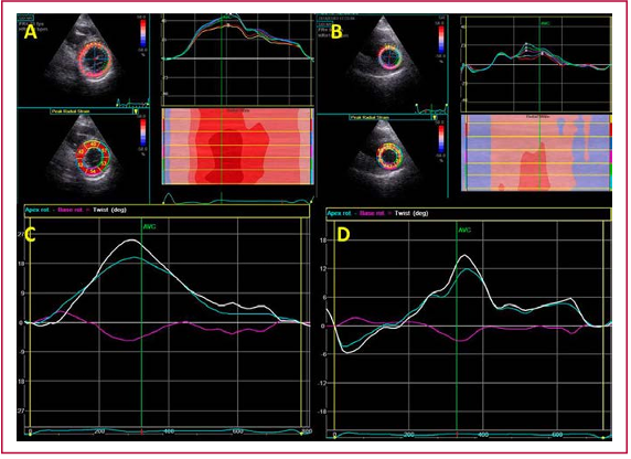
Results: Twenty-three male patients with hereditary hemochromatosis (46±18 years) and 20 male controls (45±15 years) were included. There were no differences in ventricular dimensions and wall thickness. Left ventricular ejection fraction was lower in patients with hereditary hemochromatosis (59±4% vs. 62±4%; p=0.01). There were no differences in MAPSE, TAPSE and left ventricular s’ velocity. There was a significant decrease in global strain in patients with hereditary hemochromatosis, with greater involvement of radial strain (37±12 % vs. 55±17 %; p=0.01) and circumferential strain (-19.5±2.8 % vs. -22.5±2.8 %; p=0.001), and less involvement of longitudinal strain (-19±1.8 % vs. -21.1±2.5 %; p=0.04). Myocardial rotation showed lower twist (17.7±13°vs. 25±7°; p=0.03) and lower torsion (2.3±1.8°/cm vs. 3.3±1.1 °/cm; p=0.03). There was no correlation between the different strain parameters and iron metabolism.
Conclusions: Echocardiography, and particularly 2D strain analysis can detect early abnormalities of ventricular mechanics in asymptomatic patients with systemic iron overload due to hereditary hemochromatosis.
Downloads
Published
Issue
Section
License
Copyright (c) 2019 Argentine Journal of Cardiology

This work is licensed under a Creative Commons Attribution-NonCommercial-ShareAlike 4.0 International License.