
Factors Associated with Non-calcified Plaques in Patients with Coronary Artery Calcium Score of Zero
pp. 315-321
DOI:
https://doi.org/10.7775/rac.es.v88.i4.16435Keywords:
Computed Tomography Angiography / methods - Vascular Calcification / diagnostic imaging - Plaque, Atherosclerotic / diagnostic imaging - Risk AssessmentAbstract
Background: The coronary artery calcium score is used for risk stratification in asymptomatic patients. Although coronary artery disease can occur in the absence of coronary artery calcifications, no conditions associated with the presence of soft non-calcified plaques have been described in this scenario, beyond the presence of symptoms.
Objectives: The aim of this study was to determine the associations between non-calcified plaques and independent variables in patients with coronary artery calcium score of zero.
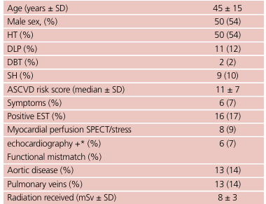
Methods: Consecutive patients with coronary artery score of zero Agatston units who also underwent computed tomography coronary angiography were included in the study. Univariate logistic regression analysis was used to find associations. (15) Sensitivity, specificity, positive predictive value (PPV), negative predictive value (NPV), positive likelihood ratio (LH+) and negative likelihood ratio (LH-) were calculated.
Results: Among a total of 93 patients, 10% (n = 9) presented non-calcified plaque. A positive exercise stress test was associated with plaques of any degree of severity (OR 6.5; 95% CI, 1.3-33, p = 0.02). This association persisted for non-severe plaques when the positive exercise stress test was combined with a negative myocardial perfusion SPECT or stress echocardiography for ischemia (OR, 12.4; 95% CI 1.5-101, p = 0.02). Sensitivity and specificity of ST-segment depression for non-calcified plaque of any degree of severity was 44.4% and 86%, respectively, with NPV of 94%, PPV of 25%, LR+ of 3.11 and LR– of 0.65.
Conclusions: ST-segment depression could be associated with non-calcified plaques in patients without coronary artery calcifica tions, even with normal exercise stress myocardial perfusion or wall motion (non-obstructive disease).
Downloads
Published
Issue
Section
License
Copyright (c) 2020 Argentine Journal of Cardiology

This work is licensed under a Creative Commons Attribution-NonCommercial-ShareAlike 4.0 International License.


