
Electrocardiographic Findings in 302 Patients in Prone position due to COVID-19
pp. 342-346
DOI:
https://doi.org/10.7775/rac.es.v89.i4.20428Keywords:
COVID-19 - SARS-CoV-2 - Electrocardiogram - Prone positionAbstract
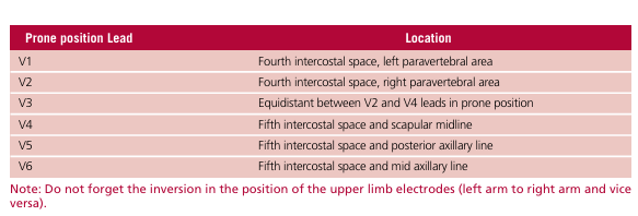
Background: Respiratory distress syndrome in patients with SARS CoV-2 poses the need for prolonged prone position. This hinders the performance of a conventional electrocardiogram (ECG), leading to consider the one obtained in prone position.
Objective: The aim of this study was to determine the electrocardiographic findings in patients in prone position and compare them with those obtained in supine position.
Methods: Patients in prone position due to respiratory distress syndrome were included in the study. An ECG was performed with definition of the most frequent findings which were compared with those observed in supine position. A p value <0.05 was considered statistically significant.
Results: A total of 302 patients in prone position showed: low voltage in 232 patients (76.8%), counter-clockwise rotation in 207 (68.5%), QS image in right precordial leads in 198 (65.6%), T wave abnormalities in 193 (63.9%), supraventricular arrhythmias in 134 (44.4%), ventricular arrhythmias in 59 (19.5%), and ischemic events in 2 (0.7%) cases.
Conclusions: The most frequent electrocardiographic findings were low voltage, counter-clockwise rotation, QS pattern in right precordial leads and reduced P wave and QRS complex voltage.
How to cite this article:
Levin R. Electrocardiographic Findings in 302 Patients in Prone position due to COVID-19. REV ARGENT CARDIOL 2021;89:342-346.
Downloads
Published
Issue
Section
License
Copyright (c) 2021 Argentine Journal of Cardiology

This work is licensed under a Creative Commons Attribution-NonCommercial-ShareAlike 4.0 International License.


