Real-Time Three-Dimensional Echocardiography
pp 365-375
DOI:
https://doi.org/10.7775/rac.v67i3.3647Keywords:
Cardiac ultrasound, Three-dimensional Echocardiography, Ventricular functionAbstract
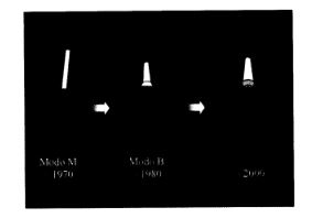
Conventional echocardiography and its represen-tation of the heart in a two-dimensional format only provide partial information about cardiac function. The development of three-dimensional echocardiography permits a more objective and quantitative assessment of cardiac physiology. Real-time three-dimensional echocardiography was developed at Duke University and is based on the design of an ultrasound transducer with a matrix array that instantaneously acquires the image contained in a three-dimensional pyramidal volume. The simultaneous visualization of multipletomographic images allows the anatomically correct examination of any structure contained within the volumetric image. Software and technologies based on high performance computers designed for graphic handling of three-dimensional images permit the rapid mapping of the volumetric image and provide possibilities beyond those of the echograph. Using this methodology, it is possible to simultaneously visualize multiple superimposed planes in a completely interactive manner controlled by the operator. Real-time three-dimensional echocardiography also allows a quantitative assessment of cardiac volumes, ventricular mass, and even myocardium with contraction and/or perfusion abnormalities. The recent addition of color flow Doppler offers other possibilities not yet explored. This technique thus expands the abilities of non-invasive cardiology and opens new doors for the diagnosis and clinical investigation of cardiac disease.
Downloads
Published
Issue
Section
License
Copyright (c) 2026 Argentine Journal of Cardiology

This work is licensed under a Creative Commons Attribution-NonCommercial-ShareAlike 4.0 International License.