

Detection of coronary artery disease is essential in the evaluation of patients with dilated cardiomyopathy and is a common practice in our setting. However, it is important to emphasize that the presence of obstructive coronary artery disease, even if extensive, does not necessarily indicate the presence of ischemic cardiomyopathy, as both conditions may coexist without a causal relationship. (1)
Bawaskar et al. recently published a registry of 3023 patients with documented obstructive coronary artery disease defined as ≥70% stenosis in ≥1 of the left anterior descending coronary artery (LAD), circumflex artery, or right coronary artery and/or ≥50% stenosis of the left main coronary artery identified on invasive coronary angiography, who underwent cardiac magnetic resonance (CMR) imaging. In this study, 9.3% of patients had non-ischemic cardiomyopathy, and this subgroup had higher risk of cardiovascular events during follow-up. (2)
STUDIES EVALUATING REVASCULARIZATION IN ISCHEMIC CARDIOMYOPATHY
In the STICH study published in 2011, patients with left ventricular ejection fraction (LVEF) less than or equal to 35% and extensive coronary artery disease amenable to coronary artery bypass grafting were randomly assigned to revascularization surgery or medical treatment, with no significant differences in death from any cause at 56 months. (3) The possible causes of the lack of response to revascularization identified in the viability sub-study were the use of methods other than CMR imaging for the assessment of viability, and the probable inclusion of patients with ventricular remodeling with a low probability of reversal, as evidenced by the high ventricular volumes recorded. (4)
In the REVIVED study published in 2022, patients with LVEF less than or equal to 35%, extensive coronary artery disease amenable to percutaneous coronary intervention and viability in at least 4 segments, were randomly assigned to percutaneous coronary intervention or optimal medical treatment with no significant differences in the composite end point of death from any cause or heart failure at 41 months. Although viability was assessed using CMR imaging in 70% of the cases, again patients with excessively high volumes were not excluded and any dysfunctional segment with less than 25% late gadolinium enhancement was considered viable. (5)
A CASE THAT POSES A DISTURBING QUESTION
In a patient with 3-vessel disease and a dilated left ventricle with global hypokinesia and severe systolic dysfunction, the absence of necrotic tissue on CMR imaging (Figure 1) suggests that the myocardium is entirely viable. However, ventricular function is unlikely to improve after revascularization because the likelihood of ischemic cardiomyopathy is very low.
Fig. 1
Images of a 64-year-old patient hospitalized for congestive heart failure. A) Coronary angiography (CA) showing three-vessel disease: severe involvement of the left anterior descending artery (LAD) , ramus intermedius (RI) and right coronary artery (RCA). B) Cardiac magnetic resonance (CMR) revealing a dilated left ventricle with global hypokinesia, normal wall thickness and absence of late gadolinium enhancement (LGE), interpreted as non-ischemic necrotic cardiomyopathy.
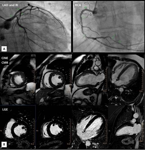
Let us now imagine that we are recruiting patients for the REVIVED study. Are there any inclusion or exclusion criteria to consider this patient not eligible? This case represents a clear example of a patient with non-ischemic cardiomyopathy who could have been included in the study.
CLARIFYING BASIC CONCEPTS
Cardiomyopathy, ischemic heart disease or dual mechanism?
By definition, cardiomyopathy is a myocardial disorder in which the heart muscle is structurally and functionally abnormal, in the absence of coronary artery disease, hypertension, valvular disease, and congenital heart disease that may explain the cardiac dysfunction. (6) It is essential to emphasize that cardiomyopathies can coexist with ischemic heart disease, valvular heart disease or hypertension and, therefore, the presence of one condition does not exclude the other. Therefore, it is essential not only to assess the presence of obstructive coronary artery disease, but also to demonstrate that it explains the extent of left ventricular dysfunction.
What do we mean when we talk about viability?
Viable myocardium is defined as myocardial tissue with reversible dysfunction caused by coronary artery disease. (7) Two main mechanisms are responsible for dysfunctional myocardium at rest with viability: stunning and hibernation. (8,9) Myocardial stunning is a phenomenon of reversible myocardial contractile dysfunction after a short period of ischemia followed by restoration of perfusion. Myocardial stunning occurs during the transient period when perfusion has normalized but myocardial contractile dysfunction persists. (10) A common example in clinical practice is the transient ventricular dysfunction that occurs after reperfusion during an acute coronary syndrome. On the other hand, the hibernating myocardium results from prolonged hypoperfusion at rest associated with myocardial metabolic demand that surpasses the coronary flow reserve. This type of myocardium presents persistent dysfunction at rest due to chronic insufficient coronary artery flow and may be partially or fully reversible following revascularization procedures. (11) A typical example of hibernation in clinical practice can be observed in chronic coronary artery occlusions with collateral circulation and minimal necrotic tissue.
Can we talk about viable myocardium in the absence of hibernation or stunning?
This question leads us to one of the conceptual errors with the greatest impact on both daily practice and the establishment of criteria for the selection of populations in randomized studies: ischemic myocardium is not usually considered in the search for viable myocardium. Ischemic myocardium is characterized by normal wall motion, metabolism and perfusion at rest; however, it becomes dysfunctional under stress conditions due to vascular impairment. If we understand this concept, it is evident that a non-necrotic myocardial segment can be ischemic. The only way to determine this is to perform a stress protocol to induce myocardial ischemia, either by stress CMR, gated SPECT or stress echocardiography.
Consequently, if the STICH and REVIVED studies did not quantify ischemia, how can we differentiate ventricular dysfunction due to coronary artery disease from cardiomyopathy with associated obstructive coronary anatomy?
Can we speak of alive but non-viable myocardium?
Hibernation and stress-induced ischemia represent different spectra of myocardial involvement due to chronic coronary artery disease. (12) Therefore, they should be considered within the spectrum of what we call clinically significant viability. In other words, a mass of myocardium worthy of revascularization. Therefore, in the context of a dilated and globally dysfunctional left ventricle, the presence of significant percentages of myocardium without necrosis, but with neither hibernation nor ischemia, indicates that we are dealing with alive (non-necrotic) but non-viable myocardium. This is because the etiology of this dysfunction is not coronary artery disease, and therefore revascularization will not improve ventricular function.
A PROPOSAL TO FACILITATE DAILY PRACTICE
According to the Argentine National Registry of Cardiac Resonance (RENAREC), viability evaluations account for approximately 5% of CMR imaging requests in our country. (13) To ensure the maximum utility of this resource, which is difficult to access in our environment, we propose the adoption of a unified terminology that is simple to implement. This terminology could help clinical cardiologists who refer patients for cardiovascular imaging tests intended to assess myocardial viability to understand this concept. We suggest referring to clinically significant viability which means hibernating or ischemic myocardium, excluding myocardium that is alive but not affected by coronary artery disease (Table 1).
Table 1
Proposal for a simplified nomenclature for viability.
| Significant necrosis | Coronary artery disease | Hibernation or significant ischemia | Improves with revascularization | |
|---|---|---|---|---|
| Clinically significant viability | NO | YES | YES | YES |
| Non-viable alive myocardium | NO | NO | NO | NO |
| Non-viable necrotic myocardium | YES | YES | NO | NO |
As previously mentioned, it is imperative to clarify that, with certain exceptions, stunning is a phenomenon predominantly associated with post-reperfusion dysfunction following acute coronary syndrome. This phenomenon is uncommon in cases of chronic coronary ventricular dysfunction. Consequently, myocardial viability in an outpatient setting primarily refers to situations of hibernation or ischemia.
FINAL COMMENT
When referring to viable myocardium or clinically significant viability, we assume that ventricular dysfunction results from coronary artery disease. As with other medical specialists, cardiologists formulate diagnostic interpretations that are subject to debate. Therefore, we propose to talk about viable myocardium only under the suspicion of underlying coronary artery disease. It is essential to have data derived from clinical examination and imaging tests that support our hypothesis of myocardial dysfunction due to obstructive coronary artery disease (causality) and that it is not a mere coincidence between the two conditions.
For this purpose, we believe that a proper diagnostic approach should include the assessment of myocardial ischemia and not be limited only to the absence of necrosis on CMR imaging with gadolinium-based contrast agent or resting myocardial perfusion scan.
We cannot claim an improvement in ventricular function after revascularization without first understanding the pathophysiology of ventricular dysfunction.
Conflicts of interest
None declared. (See authors' conflict of interests forms on the web).