

Coronary artery pseudoaneurysms are a rare but potentially fatal complication characterized by disruption of the arterial wall with flow contained either by perivascular tissues or an organized thrombus. Most pseudoaneurysms are associated with percutaneous coronary interventions, while those due to infections are rare and typically related to bacteremia or infective endocarditis caused by Staphylococcus aureus. (1,2) The left main coronary artery is rarely involved and poses a diagnostic and therapeutic challenge due to high risk of rupture, thrombosis, and sudden cardiac death, with limited evidence available to guide its management. (2,3)
We present the case of a patient with infective endocarditis following a recent percutaneous coronary intervention of the left main coronary artery, who, among other clinical complications, developed a pseudoaneurysm of the left main coronary artery, diagnosed and characterized using multimodality cardiovascular imaging.
A 68-year-old male patient with a history of hypertension, type 1 diabetes mellitus, and prostate cancer treated in 2019 was admitted for persistent fever after undergoing a percutaneous coronary intervention to the distal left main coronary artery and ostium of the left circumflex artery, with the implantation of two drug-eluting stents. After cultures were taken and intravenous antibiotics were initiated, transesophageal echocardiogram revealed a 6.3 × 3.3 mm vegetation on the anterior mitral leaflet. This finding, along with blood cultures positive for methicillin-susceptible Staphylococcus aureus, led to the diagnosis of infective endocarditis. The patient initially presented a right pseudoaneurysm of the radial artery, which was managed with surgery, and acute kidney injury requiring temporary hemodialysis. Once fever had resolved and follow-up cultures remained negative, he developed dyspnea and hemodynamic impairment. An emergency echocardiogram was performed, revealing a new cavity adjacent to the left coronary sinus, suggestive of a pseudoaneurysm, wall motion abnormalities on the anterior wall, and a marked decrease in left ventricular ejection fraction. Coronary angiography confirmed a pseudoaneurysm of the left main coronary artery with contrast agent passage into the periarterial space (Figure 1). There was also a new severe stenosis of the proximal left anterior descending coronary artery, which required implantation of a drug-eluting stent. Coronary computed tomography angiography demonstrated a partially thrombosed pseudoaneurysm extending into the proximal segments of the left anterior descending and circumflex arteries (Figure 2).
Fig. 1
Coronary angiography showing a pseudoaneurysm of the left main coronary artery with contrast agent passage into the periarterial space.
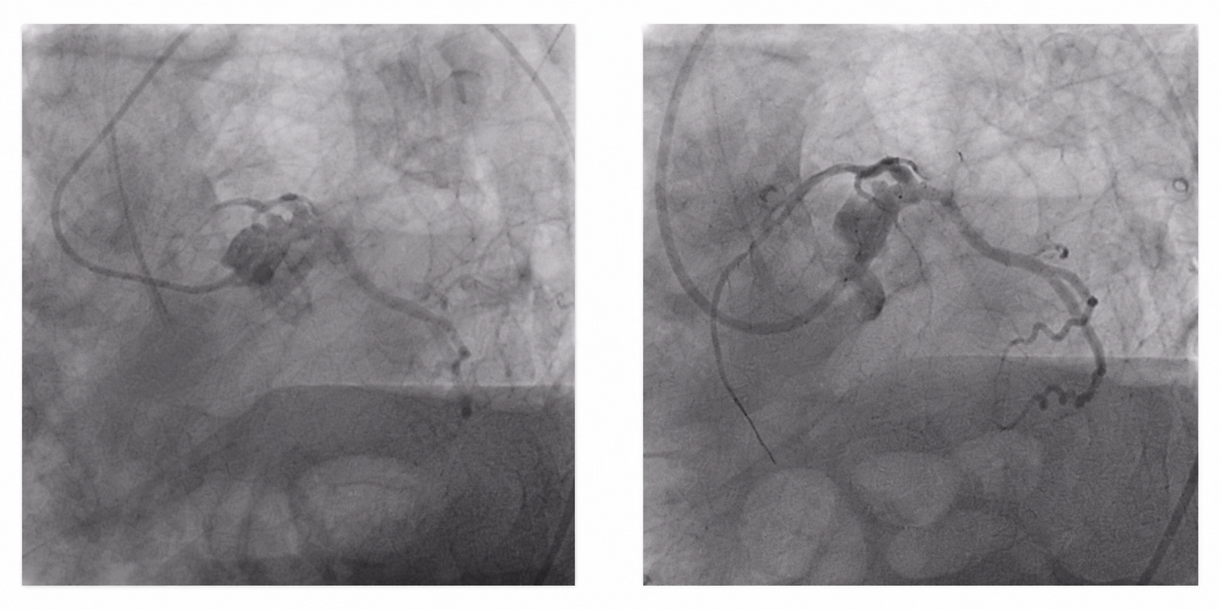
Fig. 2
Coronary computed tomography angiography confirming a partially thrombosed pseudoaneurysm extending into the proximal segments of the left anterior descending and circumflex arteries.

Given the high risk of rupture and the high surgical risk, the Heart Team decided to exclude the pseudoaneurysm by implanting a covered stent in the left main coronary artery. The patient had a favorable outcome and left ventricular function showed a partial recovery.
At the 4-month follow-up, the patient experienced recurrent chest pain, and a new coronary angiography was performed. The covered stent in the left main coronary artery was patent and the pseudoaneurysm was completely excluded, but the left anterior descending and circumflex arteries presented severe stenoses. Therefore, the Heart Team indicated elective coronary artery bypass grafting, after which the patient had a favorable postoperative course.
Coronary artery pseudoaneurysms are a rare complication following percutaneous coronary interventions. In addition to the mechanical trauma associated with such interventions, factors such as local inflammation, delayed endothelization, and infection may contribute to their development, particularly following the implantation of drug-eluting stents. (4)
In cases of infection, Staphylococcus aureus is the most common causative microorganism, and the right coronary artery is the most common location. Involvement of the left main coronary artery is rare, as demonstrated by the largest published case series. (2)
The clinical presentation is often nonspecific, and the diagnosis requires a high index of suspicion, especially in patients with persistent fever or bacteremia following a coronary artery intervention. In our case, the diagnosis did not arise from the initial infection but rather from the clinical and hemodynamic impairment, which prompted a new echocardiographic assessment. The sequential integration of echocardiography, coronary angiography, and coronary computed tomography angiography confirmed the diagnosis, defined the extent of the lesion, and allowed for treatment planning, highlighting the value of multimodality cardiovascular imaging in this type of patient.
The management of coronary artery aneurysms and pseudoaneurysms should be tailored based on etiology, anatomy, and risk of complications, given the lack of comparative studies and specific recommendations. (5) Although surgery remains the treatment of choice for mycotic pseudoaneurysms, Kilic et al. reported that drug-eluting stents may constitute an alternative for treating certain types of pseudoaneurysms when the immediate surgical risk is high, as in the case of our patient. Yet the authors warned about the increased risk of thrombosis and restenosis. (3)
In addition to reporting the exceptional location of this pseudoaneurysm, we believe the primary contribution of this case is demonstrating that a strategy based on multimodality imaging and Heart Team discussion is essential for both diagnosis and treatment planning. (6)
Ethical considerations
Not applicable