Ablación de taquicardia auricular de aneurisma de la orejuela derecha

Rev Argent Cardiol 2024;92:286-287. http://dx.doi.org/10.7775/rac.v92.i4.20806
Correspondence: Rocío B. Benito. Email: rocio.benito93@gmail.com
MTSAC Miembro Titular de la Sociedad Argentina de Cardiología

Ectopic atrial tachycardia (AT) is a relatively uncommon form of supraventricular tachycardia involving challenging medical care. Radiofrequency ablation (RFA) therapy has a high long-term success rate. (1,2)
Foci tend to be located in distinct anatomical regions. In the right atrium, these foci are found along the crista terminalis, the tricuspid annulus, the coronary sinus ostium, and the right atrial appendage (RAA). The latter is the most frequent focus in young subjects. (1,3)
RAA aneurysms are rare, and focal AT derived therefrom even more. (4) Fewer than 25 cases have been reported to date, and most of them have required surgical treatment (appendage resection). The best way to achieve a definitive diagnosis consists in using multimodal imaging. (5)
We present the case of a 25-year-old female patient with ectopic AT resulting from RAA aneurysm, refractory to medical treatment, treated with RFA.
The patient experienced sporadic palpitations and dyspnea. Holter ECG showed predominant sinus rhythm, with an average heart rate of 133 beats/minute (range: 86-179), and AT episodes. Laboratory tests and cardiac Doppler ultrasound were normal. She was initially administered ivabradine 5 mg/12 h and propanolol 80 mg/8 h. Due to the lack of response to medical treatment, both RFA and 3D mapping of the right AT were performed, guided by intracardiac echocardiography to reach the base and distal third of the RAA.
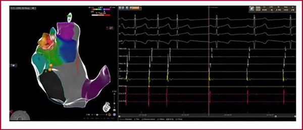
After 9 months the patient remained symptomatic and refractory to multiple medical therapies. Holter ECG revealed tachycardia of 177 bpm. A computed angiotomography showed a broad-base RAA with the upper end extending from the right atrium. A new ablation was performed. Using a CARTO 3 3D navigation system and a Pentaray® catheter, the reconstruction and activation mapping of the right atrium, superior vena cava and RAA were achieved. Angiography of the RAA (Figure 1A) showed a lobulated and wide-base RAA with a narrow-base aneurysm of about 3 cm in diameter, moved by heartbeats. Intracardiac echocardiography (Figure 1B) confirmed the anatomy and guided ablation. A precocity mapping was performed from the base of the aneurysm to the inner part, where precocity improved (-45 msec). Using a Smarttouch SF® external irrigated catheter, 5 applications were administered inside the aneurysm but they failed to stop tachycardia. We then proceeded to complete applications with a Freezor™ Xtra catheter (-80°C) for 240 seconds in that area, with no evidence of stopping tachycardia. Finally, we decided to electrically isolate the aneurysm with the RF catheter, and 8 seconds after the last application, AT stopped. (Figure 2). After a 30-minute period of observation, atrial stimulation protocol, and from the interior of the aneurysm, was performed, without tachycardia induction.
Fig. 1
A. Radioscopy showing right atrial appendage and aneurysm, with an ablation catheter inside. B. Intracardiac echocardiography from the right atrium displaying right atrial appendage and aneurysm.
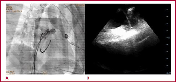
Fig. 2
Activation mapping with right atrial appendage isolation which stops atrial tachycardia during applica tions.
At the 3-month post-procedure follow-up, the patient is asymptomatic, and Holter ECG has revealed no new tachycardia occurrences.
In patients with ectopic AT, transcatheter ablation is considered the first-line therapy. Even though there is limited experience with this technique, success rates are 80-90%. In this case, we believe that successful ablation was due to the combined use of 3D mapping, intracardiac echocardiography and angiography, which allowed RF applications together with cryoablation at the site of interest to achieve electrical isolation of the aneurysm and, therefore, AT disappearance.
REFERENCES
1. Kistler PM, Roberts-Thomson KC, Haqqani HM, Fynn SP, Singarayar S, Vohra JK, et al. P-wave morphology in focal atrial tachycardia: development of an algorithm to predict the anatomic site of origin. J Am Coll Cardiol. 2006;48:1010-7. https://doi.org/10.1016/j.jacc.2006.03.058
2. Mizui S, Mori K, Kuroda Y. Ectopic atrial tachycardia due to aneurysm of the right atrial appendage. Cardiol Young. 2001;11:229-32. https://doi.org/10.1017/S1047951101000178
3. Bokhari F, Alqurashi M, Raslan O, Alama N. Right atrial appendage tachycardia: A rare cause of tachycardia induced cardiomyopathy with successful radiofrequency ablation using the 3D mapping system. J Saudi Heart Assoc. 2013;25:265-71. https://doi.org/10.1016/j.jsha.2013.04.002
4. Kanaya T, Nishigaki K, Yoshida Y, Nakamura Y, Murakami Y, Suzuki T. Ectopic atrial tachycardia originating from right atrial appendage aneurysms in children: Three case reports. Heart Rhythm Case Rep. 2017;4:2-5.https://doi.org/10.1016/j.hrcr.2017.10.010.
5. Bachani N, Jadwani J, Joshi S, Panicker GK, Lokhandwala Y. Right atrial Appendage Aneurysm with Intractable Atrial Tachycardia. IHJ Cardiovascular Case Reports (CVCR) 2019;3:108-111. https://doi.org/10.1016/j.ihjccr.2019.12.009