Riesgo de eventos embólicos en pacientes con miocardiopatía chagásica y fibrilación auricular a pesar de terapia antitrombótica: ¿es la anticoagulación suficiente

Rev Argent Cardiol 2024;92:278-285. http://dx.doi.org/10.7775/rac.v92.i4.20795
Received: 04/01/2024 Accepted: 08/15/2024
Correspondence: Luis Eduardo Echeverría Correa. E-mail: luisecheverria@fcv.org

INTRODUCTION
Chagas disease (CD) is endemic in American tropical and subtropical regions and has represented a public health problem in Latin America for decades. However, due to migration, it has become a global economic and health burden. (1) It is estimated that there are six million people infected with T. cruzi in Latin America, 300 000 in the United States, and 42 000 in Europe. (2) Chagas disease attributes a substantial burden to the patient and society regarding direct and indirect costs associated with medical care, early mortality, disability, and negative labor consequences such as loss of productivity due to absenteeism and tardiness. (3)
Chagas disease is known to have two forms: acute and chronic. (4) The acute phase of the disease is usually asymptomatic. After this phase, the patient enters a chronic phase, in which about 60-70% remain in the indeterminate form of the disease, characterized by positive serology for CD, but no evidence of structural cardiomyopathy or heart failure (HF) symptoms. (5) In this context, 30-40% of cases progress to clinical forms of the disease, with the cardiac form being the most frequent and severe manifestation. (6,7) Cardiac involvement is characterized by symptoms of HF, alterations of the conduction system, bradyarrhythmia and tachyarrhythmias, in addition to radiographic and echocardiographic alterations. (8,9) Besides, HF of chagasic origin is associated with higher mortality compared with other etiologies. (10)
During the chronic phase, multiple alterations in the structure and function of the myocardium lead not only to the development of HF symptoms but also to the appearance of prothrombotic conditions such as atrial fibrillation (AF) or even intraventricular clots, which are the product of the slow flow and structural alterations in the endocardium. (11,12) This association has been evidenced in some studies evaluating patients with cardiomyopathy of chagasic origin compared with cardiomyopathies of other etiologies. (13) This differential risk could be explained by some of the characteristics of chagasic cardiomyopathy (CC), such as a higher incidence of AF and apical aneurysms. Consequently, multiple studies have found a high incidence of ischemic stroke in patients with heart failure of chagasic etiology, highlighting the significant risk of embolic events with this condition. However, it is unknown whether there is benefit from anticoagulation in these patients despite not meeting the criteria for receiving anticoagulants by CHA2DS2-VASc scoring. Besides, few studies have compared the risk of embolic events exclusively in anticoagulated patients with CC versus other heart failure etiologies. Therefore, the objective of the present study was to evaluate the risk of embolic events and bleeding in anticoagulated patients with and without CC treated in a fourth-level specialized center in Colombia.
METHODS
Study population
A retrospective cohort study was conducted between 2014 and 2020 at the Heart Failure and Anticoagulation Clinic of a fourth-level hospital in Floridablanca, Colombia. The medical records of all patients assessed in the Anticoagulation Clinic were reviewed to assess their eligibility. Patients were included if they met the following criteria: a) age ≥18 years; b) having a diagnosis of cardiomyopathy of any etiology, and c) diagnosis of atrial fibrillation. The diagnosis of cardiomyopathy of other etiologies was based on the clinical records information, including hypertensive, ischaemic, and valvular etiologies in this group. On the other hand, we excluded patients with less than two follow-up appointments at the Anticoagulation Clinic and those with incomplete data in the electronic clinical records. A structured format was developed to extract information, which was collected retrospectively, covering sociodemographic, clinical, and laboratory variables.
Outcomes and main exposure
The outcomes of interest were time to first embolic event (including cerebrovascular events and systemic embolisms such as ischemic stroke, acute myocardial infarction of embolic origin, mesenteric ischemia of embolic origin, and other systemic embolisms of cardiogenic origin) and time to the first bleeding event (including minor or major skin, periodontal, other mucosae, intra-articular and central nervous system bleeding). Cardiomyopathy of chagasic origin was considered as the primary exposure, which was verified by registering a positive result of at least two different tests for Chagas Disease (Enzyme-Linked ImmunoSorbent Assay, haemagglutination-inhibition test, or indirect immunofluorescence test) and the presence of electrocardiographic abnormalities compatible with CC (left anterior fascicular block, right bundle branch block, atrioventricular blocks, ventricular premature beats, atrial fibrillation or flutter, heart rate ≤50 beats/min), or echocardiographic findings suggestive of myocardial involvement.
Statistical analysis
Categorical variables are presented as absolute numbers and proportions. The normality of the continuous variables was evaluated using histograms and the Shapiro-Wilk test. Variables with normal distribution are presented as means with their respective standard deviations and those with non-normal distribution are reported as medians with the corresponding interquartile range (IQR). A bivariate analysis comparing the sociodemographic and clinical characteristics according to the cardiomyopathy etiology was performed using the Chi-Square test, Fisher's exact test, Student´st test and the Mann-Whitney U test. The cumulative incidence of the events of interest was calculated with their respective 95% confidence intervals. Survival analysis was performed using the Kaplan-Meier method and adjusted Cox proportional hazard models. A value of p <0.05 was considered significant. All statistical tests were two-tailed. The analyses were performed using the statistical software STATA version 16.
Ethical considerations
This research was carried out following Resolution No. 08430 of 1993 of the Colombian Ministry of Health, which establishes the scientific, technical, and administrative standards for health research. The research protocol was submitted and approved by the Scientific Technical Committee and Research Ethics Committee of the Hospital.
RESULTS
A total of 149 anticoagulated patients with cardiomyopathy were evaluated (Figure 1). Median age was 71 (63-78) years and 30.20% were women. Median left ventricular ejection fraction was 35% (25-46) and median international normalized ratio (INR), 2.5 (2.2-3.1). Among these patients, 56.38% were treated with direct oral anticoagulants (DOACs); 55.17% had coronary artery disease, 26.21% chronic kidney disease; 28.86% had a Chagas disease diagnosis; 26.21% had history of bleeding, and 19.31% had history of embolic events. In 33.10% of cases, patients had an implanted device (pacemaker, cardioverter defibrillator, and others). Median CHA2DS2-VASc score was 4 (3-5) points and the median HAS-BLED score was 2 (2-3) points (Table 1).
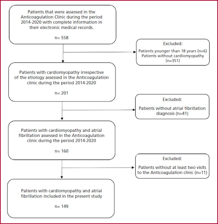
Table 1
Sociodemographic and clinical characteristics of patients with anticoagulated cardiomyopathy (n = 149).
Coronary artery disease and chronic kidney disease were more frequent in patients with cardiomyopathy of other etiologies than in patients with CC. Patients with non-chagasic cardiomyopathies had an apparent higher risk of embolisms (higher CHA2DS2-VASc score) while patients with Chagas' cardiomyopathy had a greater number of devices implanted (Table 2).
Table 2
Sociodemographic and clinical characteristics according to the cardiomyopathy etiology (n = 149)
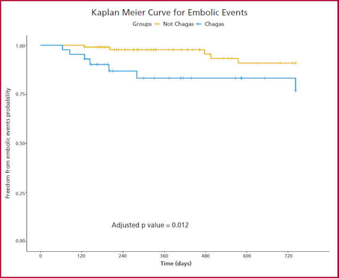
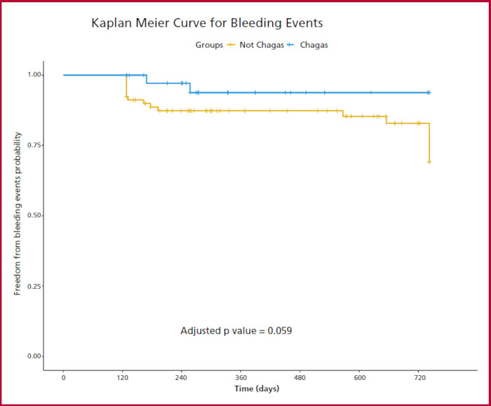
The median follow-up time for embolism was 373 (128-742) days, with a cumulative incidence of embolic events in the entire cohort of 8.05% (12/149; 95% CI 4.47-14.39%); 16.27% (7/43; 95% CI 7.34 -32.78%) in CC patients and 4.72% (5/106; 95% CI 1.63-11.17%) in other etiologies (p=0.013). Regarding bleeding, median follow-up time was 315 (117-691) days, with a cumulative incidence of 14.09% (21/149; 95% CI 9.19 - 21.28%) for the total cohort; 4.65% (2/43; 95% CI 0.58-16.16%) in CC patients and 17.92% (19/106; 95% CI 11.49-27.29%) in other etiologies (p=0.035).
Chagas disease was significantly associated with the presence of embolic events with a hazard ratio (HR) of 5.65 (95% CI 1.46-21.83; p = 0.012) in a multivariate regression model adjusted for the CHA2DS2-VASc score and the type of anticoagulant prescribed (Figure 2), but it was not associated with a higher bleeding risk: HR 0.24 (95% CI 0.05- 1.06; p = 0.059) in a model adjusted for the HAS-BLED score and the type of anticoagulant prescribed (Figure 3). For both models, the proportionality risk assumption was met.
There were no significant differences in embolic events, bleeding, and mortality according to the type of anticoagulant. (Table 3). These results were similar when analyzing only the CC population, but patients treated with warfarin in this group tended to present a higher risk of bleeding.
DISCUSSION
In the present cohort study, a significantly higher risk of embolic events was evidenced in patients with CC, despite receiving anticoagulant therapy and regardless of other clinical factors reflected by the CHA2DS2-VASc score and the type of anticoagulant prescribed. On the other hand, there was no significant difference in bleeding risk after adjusting for the HAS-BLED score and the type of anticoagulant received. These findings highlight the differential role of CC compared with other etiologies of heart failure.
Chagasic cardiomyopathy represents a unique model of thromboembolic risk due to its complex pathophysiology, which leads to a series of typical structural and functional alterations of the myocardium. (14) Some of the first evidence of the CC thromboembolic potential was reported in autopsy studies carried out almost 50 years ago, revealing that around 20% of the cases presented multiple cerebral infarcts, half of which were the cause of death. (15,16,17) Subsequently, several epidemiological studies have observed a significant association between CC and the development of symptomatic cerebrovascular disease, where the meta-analysis by Cardoso et al.¸ which evaluated eight studies that compared the risk of stroke in patients with CC in comparison to other etiologies stands out. This study concluded that patients diagnosed with CC had a risk almost two times higher for this outcome (OR 1.74, 95% CI 1.02-3.00), with a consistent result after sensitivity analysis. (13) Among the studies that compared the risk of cerebrovascular events in patients with CC versus other cardiomyopathy causes, only one corresponded to a cohort study, the majority being cross-sectional or case-control studies. (15,18,19,20,21,22,23,24)
Nevertheless, embolic events can occur even in the early stages of the disease, as reported in a cohort of patients with mild CC, in which a rate of 2.7 embolic events per 100 patient-years was estimated, being much higher than that observed in healthy patients. (25,26) However, there seems to be no difference regarding the risk of embolic events in patients in the indeterminate phase of the disease, even though a series of alterations in different coagulation factors have been documented in it, conditions which warrant further study. (25,27)
This observed high embolic risk derives not only from the development of congestive heart failure as a result of the chronic persistence of the parasite in the myocardium but also from the high frequency of severe conduction disorders (notably atrial fibrillation, intraventricular conduction defects, and even atrioventricular blocks), ventricular aneurysms, and mural thrombi, which have been significantly associated with sudden cardiac death and systemic thromboembolism in these patients. (2) However, other cardiovascular disorders observed in CC may also increase the risk of thromboembolic events, such as atherothrombosis of the great vessels and small vessel disease, which can be attributed to both typical cardiovascular risk factors and direct damage of T. cruzi to the vascular smooth muscle. (28) This may explain why about 25% of cerebrovascular events in patients with CC are classified as cryptogenic even after conducting additional studies such as echocardiography, 24-hour Holter monitoring, and carotid echo-Doppler. (29)
The present study adds relevant information to the current literature, revealing a significant difference between CC and other cardiomyopathies regarding embolic events incidence despite anticoagulation and after adjusting by relevant covariates. However, even when the potential pathophysiological mechanisms explaining these differences are well-known, there is still very poor awareness of the importance of initiating early prophylaxis in high-risk groups, although they do not fulfill the conventional criteria for anticoagulation used in other cardiomyopathies. (30,31,32) An important initiative in this aspect is being led by the Instituto de Pesquisa Evandro Chagas and the Fundação Oswaldo Cruz in Brazil. These two entities created the IPEC-FIOCRUZ score, which aims to propose prophylaxis strategies against cardioembolic ischemic stroke in Chagas disease based on clinical risk-benefit, achieving high precision in the identification of high-risk of embolic events groups (area under the ROC curve of this model was 0.90 [95% CI 0.86 - 0.94]). (33) However, the IPEC-FIOCRUZ was validated in a single center using a relatively small sample size, highlighting the need to perform additional studies to obtain an adequate external validation of this score and promote a more widespread use. This will potentially favor optimal anticoagulant prophylaxis among CC patients, further reducing the risk of embolic events in this population. (33)
Finally, not having observed differences in the risk of embolic events when comparing the types of anticoagulants used may reflect the limited statistical power of the present study derived from the relatively small sample. Currently, there is still a debate in the literature regarding the usefulness of DOAC compared to warfarin for the management of conditions such as intraventricular thrombi and anticoagulation in prosthetic valves. (34,35) There are already studies comparing the types of anticoagulants in these contexts; however, we do not have a record of a study designed to evaluate the benefit of DOAC over warfarin as anticoagulant therapy in the specific context of CD. (36,37) Given their characteristic differences and high cardioembolic risk, it is necessary to carry out research aimed at clarifying both the type of optimal anticoagulant and the particular indications for its use in these patients.
Study limitations
We must acknowledge the multiple limitations of the present study, highlighting the small sample size, which may have reduced the probability of identifying significant associations between the variables evaluated. The retrospective nature of the study may influence the quality of the information recorded. Finally, the group of cardiomyopathies of other etiologies represented a heterogeneous population, which may also interfere with the analyses performed. On the other hand, our study has a particular strength derived from the inclusion of patients only with a diagnosis of atrial fibrillation, which allowed us to demonstrate the role of other possible causes of embolism that justify the higher risk observed in the CC group.
CONCLUSIONS
Chronic CC represents a unique and characteristic myocardial involvement condition, associated with a significantly higher risk of embolic events than other cardiomyopathy etiologies, despite anticoagulation. This result was in accordance with what has been published so far in the scientific literature, with the added value of having evaluated only anticoagulated patients. It is necessary to carry out more studies that allow a clear understanding of the origin of this greater risk observed, in order to translate this knowledge into specific indications for anticoagulation in patients with CC, thus allowing an optimization in its management an improvement in its outcomes.
Financing
The present study was funded through the EMRISTA (Emerging Markets Thrombosis Investigator- Initiated Research Program) call by Pfizer, Inc. The funder had no role in the study design, data collection and analysis, publishing decision or manuscript preparation. LZR receives financial support from the Postdoctoral Stay Program of the National System of Science, Technology and Innovation, Call 848 of 2019.
REFERENCES
1. Vieira JL, Tavora FR, Sobral MG, Vasconcelos GG, Almeida GPL, Fernandes JR, et al. Chagas Cardiomyopathy in Latin America Review. Curr Cardiol Rep 2019;21:8. https://doi.org/10.1007/s11886-019-1095-y
2. Nunes MC, Beaton A, Acquatella H, Bern C, Bolger AF, Echeverría LE, et al. Chagas Cardiomyopathy: An Update of Current Clinical Knowledge and Management: A Scientific Statement From the American Heart Association. Circulation 18 de 2018;138:e169-209. https://doi.org/10.1007/s11886-019-1095-y
3. Olivera MJ, Buitrago G. Economic costs of Chagas disease in Colombia in 2017: A social perspective. Int J Infect Dis 2020;91:196-201. https://doi.org/10.1007/s11886-019-1095-y
4. Rassi A, Jr., Rassi A, Rassi SG. Predictors of mortality in chronic Chagas disease: a systematic review of observational studies. Circulation 2007;115:1101-8. https://doi.org/10.1161/CIRCULATIONAHA.106.627265
5. Prata A. Clinical and epidemiological aspects of Chagas disease. Lancet Infect Dis 2001;1:92-100. https://doi.org/10.1016/S1473-3099(01)00065-2
6. Nunes MCP, Badano LP, Marin-Neto JA, Edvardsen T, Fernandez-Golfin C, Bucciarelli-Ducci C, et al. Multimodality imaging evaluation of Chagas disease: an expert consensus of Brazilian Cardiovascular Imaging Department (DIC) and the European Association of Cardiovascular Imaging (EACVI). Eur Heart J Cardiovasc Imaging 2018;19:459-60n. https://doi.org/10.1093/ehjci/jex154
7. Lidani KCF, Bavia L, Ambrosio AR, de Messias-Reason IJ. The Complement System: A Prey of Trypanosoma cruzi. Front Microbiol 2017;8:607. https://doi.org/10.3389/fmicb.2017.00607
8. Benziger CP, do Carmo GAL, Ribeiro ALP. Chagas Cardiomyopathy: Clinical Presentation and Management in the Americas. Cardiol Clin 2017;35:31-47. https://doi.org/10.1016/j.ccl.2016.08.013
9. Rocha MO, Teixeira MM, Ribeiro AL. An update on the management of Chagas cardiomyopathy. Expert Rev Anti Infect Ther 2007;5:727-43. https://doi.org/10.1586/14787210.5.4.727
10. Rassi A, Jr., Rassi A, Little WC, Xavier SS, Rassi SG, Rassi AG, et al. development and validation of a risk score for predicting death in Chagas' heart disease. N Engl J Med 2006;355:799-808. https://doi.org/10.1056/NEJMoa053241
11. Acquatella H. Echocardiography in Chagas heart disease. Circulation 2007;115:1124-31. https://doi.org/10.1161/CIRCULATIONAHA.106.627323
12. Garcia-Alvarez A, Sitges M, Pinazo MJ, Regueiro-Cueva A, Posada E, Poyatos S, et al. Chagas cardiomyopathy: the potential of diastolic dysfunction and brain natriuretic peptide in the early identification of cardiac damage. PLoS Negl Trop Dis. 2010;4. https://doi.org/10.1371/journal.pntd.0000826
13. Cardoso RN, Macedo FYB, Garcia MN, Garcia DC, Benjo AM, Aguilar D, et al. Chagas cardiomyopathy is associated with higher incidence of stroke: a meta-analysis of observational studies. J Card Fail 2014;20:931-8. https://doi.org/10.1016/j.cardfail.2014.09.003
14. Carod-Artal FJ, Gascon J. Chagas disease and stroke. Lancet Neurol 2010;9:533-42. https://doi.org/10.1016/S1474-4422(10)70042-9
15. Lopes ER, Marquez JO, da Costa Neto B, Menezes AA, Chapadeiro ME. association of encephalic vascular accidents and Chagas disease. Rev Soc Bras Med Trop 1991;24:101-4. https://doi.org/10.1590/S0037-86821991000200006
16. Pitella JE. Ischemic cerebral changes in the chronic chagasic cardiopathy. Arq Neuropsiquiatr 1984;42:105-15. https://doi.org/10.1590/S0004-282X1984000200002
17. Aras R, Matta JAM da, Mota G, Gomes I, Melo A. Cerebral infarction in autopsies of chagasic patients with heart failure. Arq Bras Cardiol 2003;81:414-6. https://doi.org/10.1590/S0066-782X2003001200008
18. Abuhab A, Trindade E, Aulicino GB, Fujii S, Bocchi EA, Bacal F. Chagas' cardiomyopathy: The economic burden of an expensive and neglected disease. Int J Cardiol 2013;168:2375-80. https://doi.org/10.1016/j.ijcard.2013.01.262
19. Matta JAM da, Jr RA, Macedo CRB de, Cruz CG da, Netto EM. Stroke Correlates in Chagasic and Non-Chagasic Cardiomyopathies. PLOS ONE 2012;7:e35116. https://doi.org/10.1371/journal.pone.0035116
20. Leon-Sarmiento FE, Mendoza E, Torres-Hillera M, Pinto N, Prada A. J, Silva CA, et al. Trypanosoma cruzi-associated cerebrovascular disease: a case-control study in Eastern Colombia. J Neurol Sci 2004;217:61-4. https://doi.org/10.1016/j.jns.2003.08.015
21. Lima-Costa MF, Matos DL, Ribeiro ALP. Chagas disease predicts 10-year stroke mortality in community-dwelling elderly: the Bambui cohort study of aging. Stroke 2010;41:2477-82. https://doi.org/10.1161/STROKEAHA.110.588061
22. Oliveira-Filho Jamary, Viana Leila C., Vieira-de-Melo Rodrigo M., Faiçal Frederico, Torreão Jorge A., Villar Flávio A.G.A., et al. Chagas Disease Is an Independent Risk Factor for Stroke. Stroke 2005;36:2015-7. https://doi.org/10.1161/01.STR.0000177866.13451.e4
23. Paixão LC, Ribeiro AL, Valacio RA, Teixeira AL. Chagas disease: independent risk factor for stroke. Stroke 2009;40:3691-4. https://doi.org/10.1161/STROKEAHA.109.560854
24. Nunes M do CP, Barbosa MM, Ribeiro ALP, Fenelon LMA, Rocha MOC. Predictors of Mortality in Patients With Dilated Cardiomyopathy: Relevance of Chagas Disease as an Etiological Factor. Rev Esp Cardiol Engl 2010;63:788-97. https://doi.org/10.1016/S1885-5857(10)70163-8
25. Nunes MCP, Barbosa MM, Ribeiro ALP, Barbosa FBL, Rocha MOC. Ischemic cerebrovascular events in patients with Chagas cardiomyopathy: a prospective follow-up study. J Neurol Sci 2009;278:96-101. https://doi.org/10.1016/j.jns.2008.12.015
26. Bestetti R. Stroke in a hospital-derived cohort of patients with chronic Chagas' disease. Acta Cardiol 2000;55:33-8. https://doi.org/10.2143/AC.55.1.2005715
27. Pinazo M-J, Posada E de J, Izquierdo L, Tassies D, Marques A-F, Lazzari E de, et al. Altered Hypercoagulability Factors in Patients with Chronic Chagas Disease: Potential Biomarkers of Therapeutic Response. PLoS Negl Trop Dis 2016;10:e0004269. https://doi.org/10.1371/journal.pntd.0004269
28. Mannucci PM. Mechanisms, markers and management of coagulation activation. Br Med Bull 1994;50:851-70. https://doi.org/10.1093/oxfordjournals.bmb.a072930
29. Carod-Artal Francisco Javier, Vargas Antonio Pedro, Horan Thomas Anthony, Nunes Luiz Guillerme Nadal. Chagasic Cardiomyopathy Is Independently Associated With Ischemic Stroke in Chagas Disease. Stroke 2005;36:965-70. https://doi.org/10.1161/01.STR.0000163104.92943.50
31. Freitas EL, Sampaio ES, Oliveira MMC, Oliveira LH, Guimarães MSS, Pinheiro JO, et al. Atrial High-Rate Episodes and Their Association with Cerebral Ischemic Events in Chagasic Patients. Arq Bras Cardiol 2020;115:1072-9. https://doi.org/10.36660/abc.20190647
32. Matta JAM, Aras R, Macedo CRB, da Cruz CG, Netto EM. Stroke correlates in chagasic and non-chagasic cardiomyopathies. PLoS One 2012;7:e35116. https://doi.org/10.1371/journal.pone.0035116
33. Mendes FSN, Mediano MFF, Silva RS, Xavier SS, do Brasil PEA, Saraiva RM, et al. Discussing the Score of Cardioembolic Ischemic Stroke in Chagas Disease. Trop Med Infect Dis 2020;5:82. https://doi.org/10.3390/tropicalmed5020082
34. Robinson P. Long-term conditions and severe acute respiratory syndrome SARS-CoV-2 (COVID-19). Br J Community Nurs 2020;25:247-51. https://doi.org/10.12968/bjcn.2020.25.5.247
35. Abdelnabi M, Almaghraby A, Saleh Y. Will Direct Oral Anticoagulants Have a Chance in Prosthetic Valves? Eur Cardiol. 2020;15:1-2. https://doi.org/10.15420/ecr.2019.1.3.
36. Las Casas AA, Las Casas AA, Borges MA, Melo-Souza SE. Rivaroxaban for treatment of intraventricular thrombus in Chagas disease. J Cardiol Cases 2016;13:75-7. https://doi.org/10.1016/j.jccase.2015.10.010
37. Palmiero G, Melillo E, Rubino AS. "A Tale of Two Cities": Anticoagulation Management in Patients with Atrial Fibrillation and Prosthetic Valves in the Era of Direct Oral Anticoagulants. Medicina (Kaunas) 2019;55:437.https://doi.org/10.3390/medicina55080437