Síndrome de Marfan con aneurisma subclavio bilateral asociado a coartación aórtica

Rev Argent Cardiol 2024;92:291-292. http://dx.doi.org/10.7775/rac.v92.i4.20805
Correspondence: Nidhessy Pagés Morales. E-mail: nidhepages@gmail.com

Marfan syndrome is an inherited disease described in children and adults that affects the connective tissue of multiple organs and systems, such as the cardiovascular and musculoskeletal systems, eyes and skin. (1) It involves autosomal dominant inheritance due to a genetic alteration in chromosome 15; prevalence is estimated at 1 in 5000-10 000 newborns, and it affects both sexes equally. (2) Coarctation of the aorta is a highly complex and life-threatening cardiovascular malformation characterized by narrowing of the descending aorta, which in some cases may be a long hypoplastic aortic segment. It is usually located at the ductus arteriosus insertion point, distal to the left subclavian artery. It tends to be associated with anomalies in the aortic arch and results in left ventricular pressure overload. (3)
We present the case of a 51-year-old female patient with a history of arterial hypertension, ischemic heart disease and Marfan syndrome, who presents with increased soft tissue volume in the left anterior thorax. During anamnesis, she denies any pain or any other symptoms.
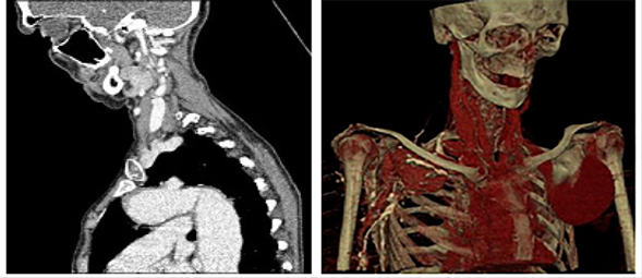
A computed tomography (CT) of the chest is performed with contrast agent using CT angiography technique, with axial slices, and both sagittal and coronal reconstructions. Aneurysmal dilation of both subclavian arteries is observed in the supraclavicular region (Figures 1 and 2). The left subclavian artery is more prominent, with an 81-mm transverse diameter and a 95-mm longitudinal diameter, and a thrombus on the wall leaving a 41-mm lumen. The contralateral artery has a 57-mm longitudinal diameter and a 36-mm transverse diameter, with absence of thrombi. The ascending aorta is dilated (53-mm), showing coarctation after the left subclavian artery emergency (Figure 2).
Figure 1
Computed tomography. A. Axial slice showing both vascular dilations at the subclavian arteries level. B. Axial slice showing aneurysmal dilation of the left subclavian artery, with both dilation and coarctation of the aorta. C. Coronal slice showing aneurysmal dilation of both subclavian arteries, the left one with a thrombus on the wall.
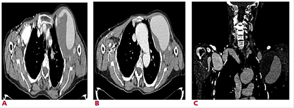
Figure 2
Computed tomography. A. Sagittal section showing dilation and coarctation of the aorta. B. Volumetric reconstruction showing both aneurysmal dilations of the subclavian arteries.
In Marfan syndrome, dilated aortic root is the most common cardiovascular occurrence, estimated to affect 60-80% of patients and to cause 90% of deaths. (4)
Aneurysms of the subclavian artery are extremely rare, with an incidence ranging from 0.01% to 3.5% as reported by different authors. It results from infections or degeneration of the tunica media of the artery, but it may be part of a clinical spectrum of diseases, including Marfan syndrome. (5) Other causes of subclavian artery aneurysm are thoracic outlet syndrome and trauma secondary to gunshot wound, clavicle fracture or iatrogenic causes. (5,6)
Complications depend on the location, and the most commonly reported are brachial plexus injury or compressed upper extremity vessels possibly leading to edema. Surgical procedures have been reported for treatment, with associated complications in almost one third of cases. (6)
REFERENCES
3. Lizano MJ, Álvarez Urbina J. Coartación de aorta congénita: una actualización en el abordaje diagnóstico y terapéutico. Revista Ciencia Y Salud Integrando Conocimientos. 2023 7(2). https://doi.org/10.34192/cienciaysalud.v7i2.617
4. Michel-Martínez AJ, Chavez-Camacho RJ, Laguna-Chavez RA. Compromiso cardiovascular en el síndrome de marfan: reporte de caso: Rev Cient Cienc Med 2020;23:252-7. https://doi.org/10.51581/rccm.v23i2.18
5. Trujillo-Santos AJ, Jurado Gámez B. Aneurisma de la arteria subclavia izquierda: causa infrecuente de masa pulmonar superior. Ann Med Inter (Madrid) [Internet]. 2001;18:40-1. https://doi.org/10.4321/S0212-71992001000200007
6. Peña-Juárez RA, Medina-Andrade MA, Ramírez-Cedillo D, Landeros A, Abarca-De la Paz JP. Aneurisma subclavio congénito en un paciente pediátrico. Arch Cardiol Mex 2021;91:514-5. https://doi.org/10.24875/ACM.20000543