Presencia de disautonomía como predictor del desarrollo de cardiopatía estructural en pacientes con enfermedad de Chagas

Rev Argent Cardiol 2024;92:333-344. http://dx.doi.org/10.7775/rac.v92.i5.20814
Received: 04/30/2024 Accepted: 09/02/2024
Correspondence: Daniel Agustín Chirino Navarta. E-Mail: daniel.chirino@hotmail.com

Introduction
Chagas disease (ChD) remains an important health issue in Latin America and in recent decades it has also spread to other regions including the United States and Europe due to migratory processes. In Argentina, it is estimated to affect between 1.5 and 2 million people, with between 350 000 and 500 000 suffering from some degree of heart disease. (1,2)
The chronic stage of ChD has a variable period in which there is no evidence of structural heart disease or involvement of other organ. This period is known as indeterminate or chronic ChD without overt structural heart disease. (3,4) However, about 30% of patients with positive serology will develop some type of structural heart disease along the course of the disease. In these patients, risk assessment represents an important challenge.
Several studies have demonstrated the presence of autonomic dysfunction in early stages of ChD. (5,6,7,8) These abnormalities could be involved in the progression of the disease.
The Valsalva maneuver (VM) has been widely used to assess autonomic function in many conditions. (9) The Valsalva ratio (VR), which is the ratio of maximal to minimal heart during the VM, is one of the most widely used tests for assessing autonomic function, because it is simple, reproducible and inexpensive. (9,10)
The role of dysautonomia assessed by the VR as a predictor of early development of structural heart disease in ChD patients without overt heart disease has not been well studied.
The aim of this study was to evaluate whether the VR, as an expression of dysautonomia, can predict early development of structural cardiac abnormalities during the follow-up of patients with ChD.
Methods
We conducted a prospective study in a single center with a follow-up program for patients with ChD. Between July 2016 and July 2018, patients with at least two positive serology tests for Trypanosoma cruzi (indirect hemagglutination, ELISA, or indirect immunofluorescence) were included in the study. A medical record was taken to all the patients, including physical examination and electrocardiogram (ECG), Doppler echocardiography and 24-hour Holter monitoring.
To be included, patients should not have overt heart disease, defined as absence of cardiovascular symptoms or signs, normal cardiovascular physical examination, normal ECG, echocardiogram without significant abnormalities and 24-hour Holter monitoring within normal parameters. Patients with a history of hypertension, diabetes mellitus, thyroid disease, ischemic heart disease, neurologic disease or kidney failure were excluded.
Patients who met the inclusion criteria were asked to perform the standard VM. The standard procedure is as follows: the patient lies in the supine position and is asked to blow through a mouthpiece connected to a manometer until reaching an expiratory pressure of 40 mm Hg. Once this pressure is reached, the patient maintains it for 15 seconds under constant supervision. Then, the mouthpiece is released and the patient continues breathing normally. An ECG is continuously recorded throughout the procedure, starting 15 seconds before expiration and continuing for 15 seconds thereafter.
The VR was calculated by measuring (in milliseconds) the shortest RR interval during the maneuver (denominator) and the longest RR interval shortly after the maneuver (numerator). Thus, the formula is: longest RR interval shortly after the maneuver/shortest RR interval during the maneuver = VR (Valsalva ratio). A VR < 1.1 was considered an abnormal result.
Patients were monitored periodically with scheduled medical interviews according to the institutional ChD follow-up program based on medical criteria.
The primary end point was the development of structural heart disease, defined as any of the following: left ventricular (LV) dilatation (LV diastolic diameter > 60 mm in men or > 55 mm in women), LV dysfunction [LV ejection fraction (EF) drop > 10 points or LVEF < 50%), complex ventricular arrhythmia or second-degree atrioventricular block type Mobitz II or greater.
The study was approved by the institutional review board and all the patients signed an informed consent form before being included.
Statistical analysis
Continuous variables were expressed as mean ± standard deviation and categorical variables as percentage. Continuous variables with normal distribution were compared using the Student's t test and the Wilcoxon test was used for variables with skewed distributions. Categorical variables were compared using the chi-square test and Fisher's exact test if any of the variables had an expected frequency < 5.
The Cox proportional hazards model was used for univariate analysis of the end point of development of structural heart disease. The VR was included as a continuous variable and as dichotomous variable (normal or abnormal). Those variables that resulted in significant differences between the groups with and without dysautonomia were also included. Predictor variables with a p value < 0.10 on the univariate analysis were included in a multivariate model. A survival analysis was performed using the Kaplan-Meier method for the VR dichotomized into normal/abnormal.
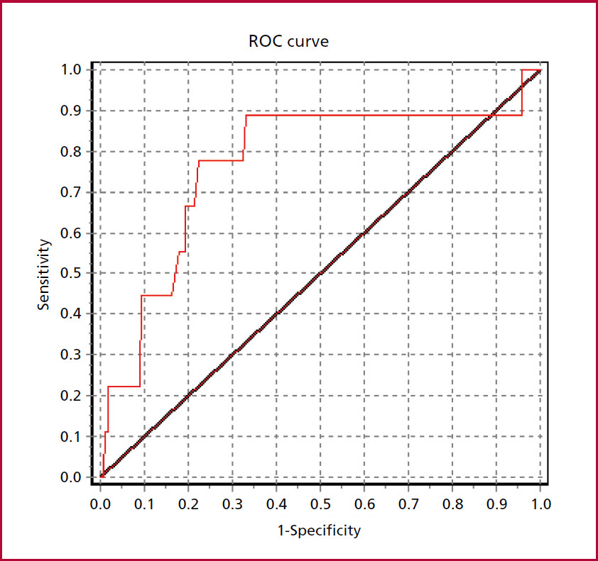
A ROC curve was constructed to establish the area under the curve (AUC) and evaluate the discriminative capacity of the VR. A p value < 0.05 was considered statistically significant. All calculations were performed using Epi-Info 7 and Statistix 8 software packages. Epidat 3.1 was used to construct the ROC curve.
Results
Of 280 patients evaluated, 80 were excluded for presenting any of the exclusion criteria. A total of 200 patients were included; mean age was 45 ± 8 years and 44% were women. Table 1 shows the baseline characteristics of the population. The VR was 1.22 ± 0.12 and was abnormal in 24% of the patients (n = 48).
Table 1
Baseline characteristics
The abnormal VR group had higher E/e' ratio on the Doppler echocardiogram (9.48 ± 2.5 vs. 7.1 ± 1.8, p <0.001) and lower S-wave in tissue Doppler imaging (0.08 ± 0.02 vs. 0.10 ± 0.02 cm/s, p = 0.023). In addition, there was trend at the limit of significance in baseline diastolic blood pressure (DBP) and maximum heart rate (HR) reached in the baseline 24-h Holter monitoring. The rest of the variables had no differences between the groups.
Median follow-up was 38 months (interquartile range 19 - 43) and the end point of development of structural heart disease occurred in 4.5% (n=9). Of these, 3 patients had increased diastolic diameter without ventricular dysfunction, LVEF decreased in 1, 4 had ventricular arrhythmia and 1 patient had second-degree AV block. There were no deaths during follow-up and only 1 patient was hospitalized for a scheduled pacemaker implantation.
Table 2 shows the univariate and multivariate analysis for the primary end point.
Table 2
Univariate and multivariate analysis for the primary end point
On multivariate analysis, both VR as a continuous variable (HR 1.02, 95 % CI 1.01-1.06, p = 0.042) and abnormal VR (HR 3.81, 95 % CI 2.81- 5.92; p=0.011) were independent predictors of development of heart disease. Figure 1 shows the Kaplan-Meier curve and the ROC curve. The area under the ROC curve was 0.77 (95% CI 0.62- 0.93) (Figure 2).
Figure 1
Kaplan-Meier curve: normal/abnormal VR for the primary end point of development of structural heart disease
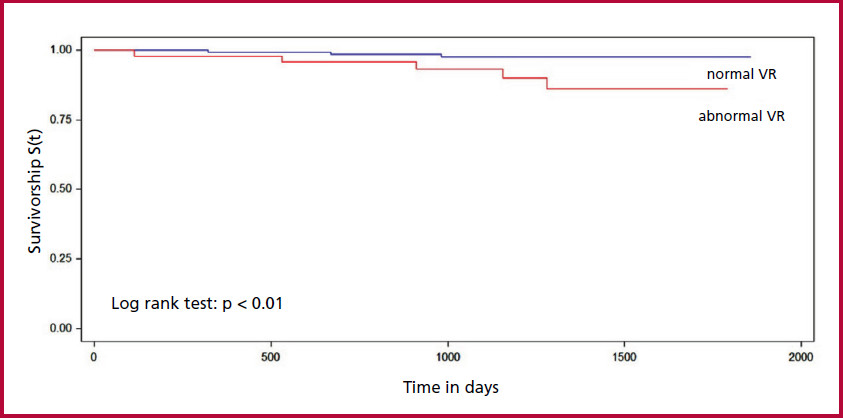
VR: Valsalva ratio
Discussion
In our study, we found that in a population of patients with ChD without overt heart disease, an abnormal VR was associated with early development of structural heart disease at a relatively short follow-up period of 3 years. Patients with an abnormal VR had a more than 3.5-fold increased likelihood of developing early heart disease, with moderate discrimination ability (AUC 0.77).
Over 20 years ago, Olivera et al. (11) found a reduction in VR among patients with ChD without overt cardiovascular disease compared to healthy controls. In addition, during follow-up, abnormal VR was associated with the development of second-degree AV block in ChD patients. Later, Ribeiro et al. (12) published a meta-analysis of 7 studies with more than 350 patients (including the study by Olivera previously mentioned) and found that the VR was decreased in patients with ChD without overt structural heart disease compared to patients without ChD.
The VR assesses the integrity of cardiac autonomic function based on heart rate responses associated with the baroreflex mechanism which is responsible for stabilizing blood pressure. (9) It is a simple, reproducible, inexpensive method that does not require major equipment. However, it is important to note that the VM involves a complex mechanism of autonomic regulation which encompasses the sympathetic and parasympathetic nervous systems. During the maneuver, blood pressure and bradycardia occur simultaneously, while at the end of the test blood pressure decreases and heart rate increases. Thus, a normal VR reflects an adequate baroreceptor response to the rise and fall of blood pressure, while a reduced VR reflects vasovagal dysfunction. (13,14)
Traditionally, a VR > 1.5 has been considered normal. (10) However, some studies have considered an abnormal VR when it is ≤ 1.1. (15,16,17) We decided to use this latter cut-off point because it is more specific of autonomic dysfunction. (17)
Impaired autonomic function has also been demonstrated in early stages of ChD compared to controls using other methods, such as heart rate variability, respiratory sinus arrhythmia magnitude (18,19) or chronotropic incompetence during exercise stress test. (19) These abnormalities occur early in the course of the disease and before the development of left ventricular dysfunction. (20) However, these tests have not evaluated whether the presence of dysautonomia predicts progression to ChD. Recently, a small study found a higher rate of exercise-induced ventricular arrhythmias and autonomic dysfunction in ChD patients without overt heart disease compared to controls without ChD. (21) Moraes et al. analyzed a cohort of 550 elderly patients with positive serology for ChD in Brazil, followed-up for 14years and found that the presence of dysautonomia was not independently associated with sudden death after adjusting for age and cardiovascular risk factors. (22)
The pathogenesis of vagal dysfunction remains unclear. Some authors have described denervation related to parasympathetic neuroganglionar lesions probably due to inflammation. (23) The role of circulating autoantibodies against muscarinic receptors that could cause desensitization and/or downregulation of these receptors has also been studied. (8) Several studies have shown increased activity of anti-M2 and anti-β1 antibodies in ChD patients with evidence of dysautonomia but with preserved ventricular function. (5)
In the present study, we found that progression to structural heart disease was 4.5% in slightly more than 3 years, which is about 1.4% per year. A recently published meta-analysis that included 23 studies found an average progression rate of 1.9% per year in patients with indeterminate ChD. This review included studies conducted in Argentina and Brazil more than 10 years ago. (24) More recently, a cohort of patients with indeterminate ChD showed a progression rate of 1.48% per year. (25) Notably, the events were mild, and no patients died during follow-up. This is similar to the systematic review by Gonzalez et al. , which revealed a remarkably low incidence of sudden cardiac death in patients with ChD without overt disease. (26)
Study limitations
Our study has some limitations. First, it is a single-center study, with a relatively short follow-up period for the type of disease. Although the progression rate was similar to the one reported, the small number of events limits the analysis. Dysautonomia was assessed using a single method, the VR. Most studies used more than one method to assess dysautonomia. We decided to use the VR because it is inexpensive, easy to perform and quite feasible to use in daily practice.
Conclusion
An abnormal baseline VR is a predictor of early development of structural heart disease in patients with ChD considered to be stage 0, with moderate discrimination ability.
Conflicts of interest
None declared. (See authors' conflict of interests forms on the web).
REFERENCES
2. Spillmann C, Burrone S, Coto HB. Análisis de la situación epidemiológica de la enfermedad de chagas en argentina: avances en el control. Rev Argent Salud Pública 2012. disponible en: https://rasp.msal.gov.ar/index.php/rasp/article/view/306/244
5. Thiers CA, Barbosa JL, Pereira Bde B, Nascimento EM, Nascimento JH, Medei EH et al. Autonomic dysfunction and anti-M2 and anti-β1 receptor antibodies in Chagas disease patients. Arq Bras Cardiol 2012;99:732-9. https://doi.org/10.1590/S0066-782X2012005000067
6. Junqueira LF Jr, Soares JD. Impaired autonomic control of heart interval changes to Valsalva manoeuvre in Chagas' disease without overt manifestation. Auton Neurosci. 2002;97:59-67.https://doi.org/10.1016/S1566-0702(02)00014-0
7. Ribeiro AL, Moraes RS, Ribeiro JP, Ferlin EL, Torres RM, Oliveira E, et al. Parasympathetic dysautonomia precedes left ventricular systolic dysfunction in Chagas disease. Am Heart J 2001;14:260-5. https://doi.org/10.1067/mhj.2001.111406
8. Ribeiro AL, Gimenez LE, Hernandez CC, de Carvalho AC, Teixeira MM, Guedes VC et al. Early occurrence of anti-muscarinic autoantibodies and abnormal vagal modulation in Chagas disease. Int J Cardiol 2007;117:59-63. https://doi.org/10.1016/j.ijcard.2006.04.053
9. Junqueira EJ. Teaching cardiac autonomic function dynamics employing the Valsalva (Valsalva-Weber) maneuver. Adv Physiol Educ 2008;32:100-6. https://doi.org/10.1152/advan.00057.2007
10. Levin AB. A simple test of cardiac function based upon the heart rate changes induced by the Valsalva maneuver. Am J Cardiol 1996;18:90-9. https://doi.org/10.1016/0002-9149(66)90200-1
11. Oliveira E, Ribeiro AL, Assis Silva F, Torres RM, Rocha MO. The Valsalva maneuver in Chagas disease patients without cardiopathy. Int J Cardiol 2002;82:49-54. https://doi.org/10.1016/S0167-5273(01)00599-X
12. Ribeiro AL, Campos MS, Baptista LM, de Sousa MR. The Valsalva maneuver in Chagas disease patients without cardiopathy. Clin Auton Res 2009;20:79-83. https://doi.org/10.1007/s10286-009-0044-z
13. Loni SBA. Valsalva ratio: a measure of stress in first year medical students. Int J Res Med Sci 2018;7:1599-604. https://doi.org/10.18203/2320-6012.ijrms20150236
14. Palamarchuk IS, Baker J, Kimpinski K. The utility of Valsalva maneuver in the diagnoses of orthostatic disorders. Am J Physiol Regul Integr Comp Physiol 2016;310:243-52. https://doi.org/10.1152/ajpregu.00290.2015
16. Pafili K, Trypsianis G, Papazoglou D, Maltezos E, Papanas N. Simplified Diagnosis of Cardiovascular Autonomic Neuropathy in Type 2 Diabetes Using Ewing's Battery. Rev Diabet Stud 2015; 12:213-9. https://doi.org/10.1900/RDS.2015.12.213
17. Ewing DJ, Martyn CN, Young RJ, Clarke BF. The value of cardiovascular autonomic function tests: 10 years experience in diabetes. Diabetes Care 1985;5:491-8. https://doi.org/10.2337/diacare.8.5.491
19. Rocha AL, Lombardi F, da Costa Rocha MO, Barros MV, Val Barros Vda C, Reis AM, et al. Chronotropic incompetence and abnormal autonomic modulation in ambulatory Chagas disease patients. Ann Noninvasive Electrocardiol 2006;3:3-11. https://doi.org/10.1111/j.1542-474X.2006.00054.x
20. Villar JC, Leon H, Morillo CAL. Cardiovascular autonomic function testing in asymptomatic T. cruzi carriers: a sensitive method to identify subclinical Chagas’ disease. Int J Cardiol 2004;93;189-95. https://doi.org/10.1016/j.ijcard.2003.03.002
21. Costa HS, Nunes MC, Souza AC, Lima MM, Carneiro RB, Sousa GR, et al. Exercise-induced ventricular arrhythmias and vagal dysfunction in Chagas disease patients with no apparent cardiac involvement. Rev Soc Bras Med Trop 2015;48:175-80. https://doi.org/10.1590/0037-8682-0295-2014
22. Moraes DN, Nascimento BR, Ribeiro AL. Abstract 11419: Prognostic Value of Vagal Dysautonomia in Patients With Chagas Disease: 14-year Results of a Population Cohort of the Elderly. Circulation 2022;(suppl146): A11419. https://doi.org/10.1161/circ.146.suppl_1.11419
23. Junqueira Júnior LF, Beraldo PS, Chapadeiro E, Jesus PC. Cardiac autonomic dysfunction and neuroganglionitis in a rat model of chronic Chagas’ disease. Cardiovascular Res 1992;26:324-9. https://doi.org/10.1093/cvr/26.4.324
24. Chadalawada S, Sillau S, Archuleta S, Mundo W, Bandali M, Parra-Henao G, et al. Risk of chronic cardiomyopathy among patients with the acute phase or indeterminate form of Chagas disease: a systematic review and meta-analysis. JAMA 2020;3:e2015072. https://doi.org/10.1001/jamanetworkopen.2020.15072
25. Hasslocher-Moreno AM, Xavier SS, Saraiva RM, Sangenis LHC, Holanda MT, Veloso HH et al. Progression rate from the indeterminate form to the cardiac form in patients with chronic Chagas disease: twenty-two-year follow-up in a Brazilian urban cohort. Trop Med Infect Dis 2020;5:E76. https://doi.org/10.3390/tropicalmed5020076
26. Gonzalez G, Ortiz Fragola J, Azzato F, Milei J. La muerte súbita no es frecuente en la enfermedad de Chagas crónica sin cardiopatía demostrable. Rev Argent Cardiol. 2019;87:53-6. http://dx.doi.org/10.7775/rac.es.v87.i1.14511