

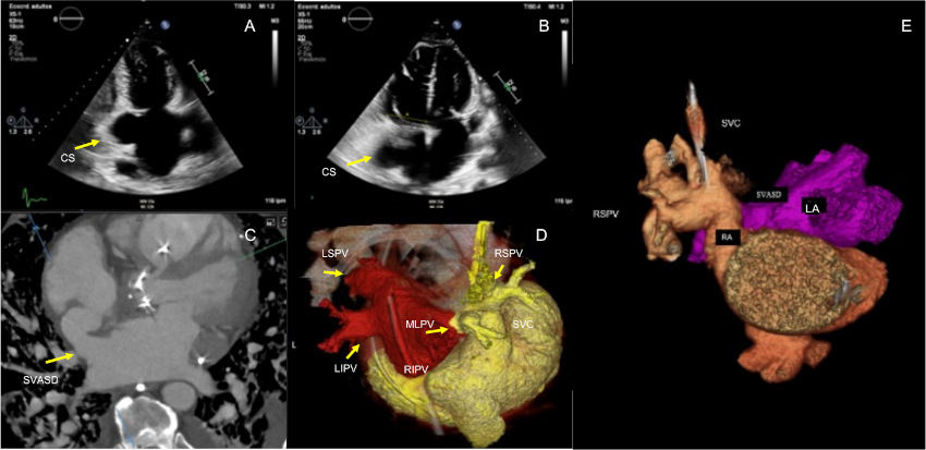
A 69-year-old male with a history of bicuspid aortic valve replacement presented with progressive right-sided heart failure. Transthoracic echocardiography revealed severe right heart enlargement and dysfunction with suspicion of an interatrial shunt (Fig. 1A-B). Transesophageal echocardiography (TEE) identified a superior sinus venosus atrial septal defect (SVASD), coronary sinus dilatation, and a persistent left superior vena cava (PLSVC), with absence of right superior pulmonary venous drainage into the left atrium.
Cardiac computed tomography (CT) confirmed the SVASD and revealed a PLSVC draining into a dilated coronary sinus, with partial anomalous pulmonary venous drainage (PAPVD) of the right superior pulmonary vein and an accessory middle lobe vein draining into the superior vena cava (Fig. 1C-E). Dilatation of the pulmonary trunk was also noted, consistent with pulmonary hypertension.
The coexistence of SVASD, PAPVD, PLSVC, and bicuspid aortic valve is exceptionally rare, with no previous reports describing this combination. SVASD is frequently associated with venous anomalies such as PAPVD and PLSVC. (1,2) This unique constellation of congenital defects highlights how one anomaly often coexists with others and underscores the pivotal role of multimodality imaging in accurate diagnosis and preoperative planning. (3)
Fig. 1
Transthoracic echocardiogram showing a dilated coronary sinus (CS) (A) and marked right ventricular enlargement (RV) (B). Cardiac CT demonstrating a superior sinus venosus atrial septal defect (SVASD) (C). Three-dimensional cardiac CT reconstruction showing the left superior (LSPV), left inferior (LIPV), and right inferior (RIPV) pulmonary veins draining into the left atrium (LA), whereas the right superior pulmonary vein (RSPV) and the accessory middle lobe pulmonary vein (MLPV) drain anomalously into the superior vena cava (SVC) (D-E).
Ethical considerations
The authors obtained informed consent from the patient.
Conflicts of interest
None declared.
(See authors' conflicts of interest forms on the website).